Guest: Albert Cheung, MD
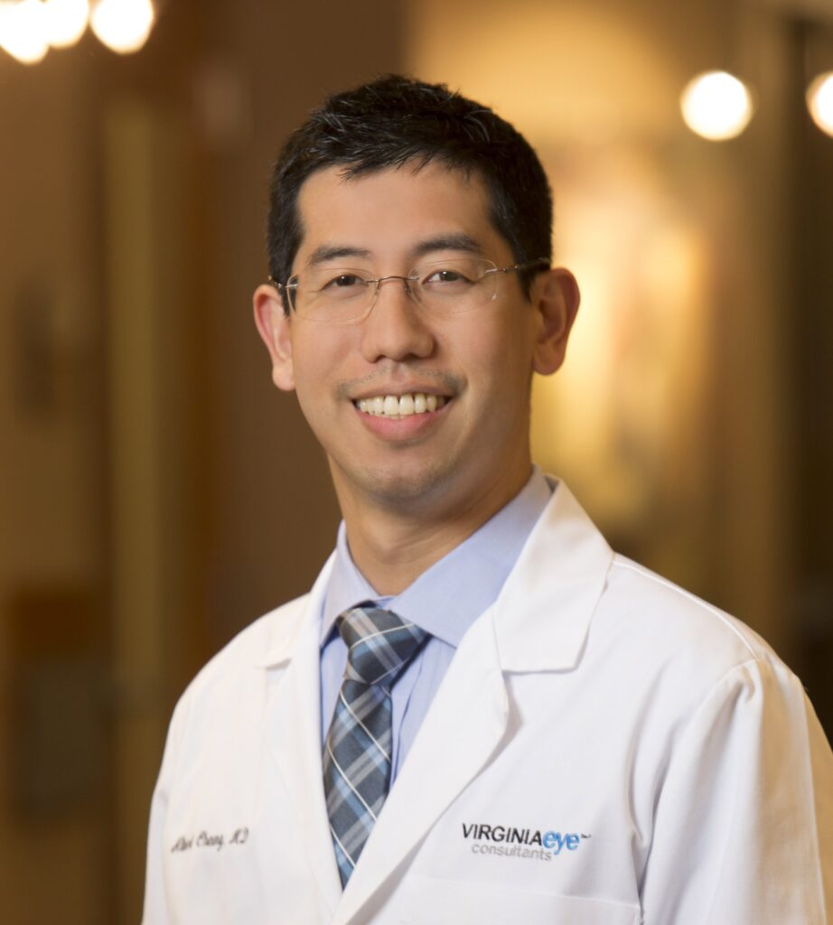
Dr. Albert Cheung is part of the skilled Cornea team at Virginia Eye Consultants in Norfolk, VA. Dr. Cheung joined VEC to provide world-class corneal and cataract care to patients. He offers advanced medical and surgical options for corneal diseases such as Fuchs dystrophy, Keratoconus, and severe ocular surface disease. He is also part of the community faculty in the Ophthalmology Department at Eastern Virginia Medical School at Old Dominion University, where Dr. Peter Netland is Chair.
Dr. Cheung has given national and international lectures on various corneal and cataract topics. As a researcher, he has authored more than 120 peer-reviewed journal articles and book chapters. Additionally, he has presented numerous papers, abstracts, and posters at national meetings. His research focus has encompassed lamellar corneal transplantation (DMEK, DSAEK, DALK), limbal stem cell deficiency, ocular surface stem cell transplantation, and cataract outcomes.
Dr. Cheung completed a Cornea/External Disease Fellowship at Cincinnati Eye Institute under Dr. Edward Holland, and he is currently working with the Holland Foundation for Sight Restoration to establish a Center of Excellence for the treatment of severe ocular surface failure.
Dr. Cheung enjoys spending his free time with family and friends. Outside of his professional career, Dr. Cheung is an experienced swimmer and has participated on varsity swimming and water polo teams. Other interests include running, drawing and painting, and following sports.
Host: Janelle Collins
Janelle serves as a Board Member at Large for ANA. She is also the Chair of the Patient and Family Advisory Board and serves as the host for Focal Point. Janelle has been involved in the aniridia patient community via both patient advocacy groups and Facebook groups for a number of years. She enjoys reading peer-reviewed journal articles and summarizing their contents for the patient community. She also is passionate about welcoming new parents of children with aniridia to the community and assisting them as they begin their aniridia journey.
Focus: The Aniridic Cornea – Treatment
Date: April 3, 2025
Time: 8:30 pm EDT
Transcript
JANELLE COLLINS:
Good evening, everyone. Welcome to the fifth episode of Focal Point. We’re still admitting the last few people who are coming, so as soon as that slows down a little bit, we will get started. I’m so glad everybody could join us.
My name is Janelle Collins, and I’m your host for tonight. I serve as the member-at-large on the board of directors of ANA, and I live in Florida in the United States with my husband and my two children. My awesome 16-year-old daughter has aniridia.
Our episode tonight is focused on the aniridic cornea. And specifically, we’re going to talk tonight about treatments. Our guest for tonight is Dr. Albert Cheung, and I’m going to go ahead and read his bio because, as usual, I did not get a chance to memorize this whole thing.
Dr. Cheung is part of the skilled cornea team at Virginia Eye Consultants in Norfolk, Virginia. He joined VEC to provide world-class corneal and cataract care to patients. He offers advanced medical and surgical options for corneal diseases such as keratoconus and severe ocular surface disease, including things like aniridic keratopathy.
He is also part of the community faculty in the ophthalmology department at Eastern Virginia Medical School at Old Dominion University, where Dr. Peter Netland is chair. Dr. Cheung has given national and international lectures on various corneal and cataract topics. As a researcher, I understand he has authored more than 120 peer-reviewed journal articles and book chapters. And additionally, he has presented numerous papers, abstracts, and posters at national meetings.
His research focus has encompassed lamellar corneal transplantation, limbal stem cell deficiency, ocular surface stem cell transplantation, and cataract outcomes.
Dr. Cheung completed a cornea and external disease fellowship at Cincinnati Eye Institute under Dr. Edward Holland, which is a name a lot of you recognize. And he’s currently working with the Holland Foundation for Sight Restoration to establish a center of excellence for the treatment of severe ocular surface failure.
Now, since I haven’t had the honor of seeing Dr. Cheung as a clinician, I asked Michael Schain, who is ANA’s newest board member, for his recent experiences.
He said “Dr. Cheung was very thorough with his initial evaluation and his presentation of my procedure. He discussed the potential upsides as well as the potential concerns. He was very generous with his time, his care, and he answered all of my questions. I know that I’m in excellent hands, and I’m confident with his guidance.”
Dr. Cheung, I am so honored that you have joined us here tonight.
DR. CHEUNG:
Well, thank you so much for having me. Thanks for the kind introduction. It’s a pleasure to be here and talk to you all.
JANELLE COLLINS:
We’re so glad to have you here. Thank you to everybody who is online who submitted questions ahead of time. Some of you submitted them for the last episode. Some of you submitted them this time.
If you think of more questions as we talk, please don’t hesitate to ask them in the chat. We are going to be trying to monitor that as we go here.
Let’s see here. I would like to show you all something. When we discussed this at the last Focal Point, we said that the cornea is just a really, really complex topic. And because it is such a complex topic, what we see as patients is often very different than what the researchers and clinicians see. We usually only see the very tip of the iceberg here, the stuff that sees the light of day. I’m hoping you all can see my iceberg picture.
But in order to get this tip of the iceberg, we have so much under the water that has to happen—that that iceberg is built on. And so last time we talked with Dr. Freeman just about that very tip. We talked about an overview of the cornea, an overview of the treatments.
Tonight, Dr. Chung and I are going to talk even more about the clinical care and the treatments, and then also about clinical research and some of the treatments that are coming up.
So just know that this is a very deep topic. And so if we don’t get to all of your questions tonight, or if your question falls maybe into one of these lower levels, we will do our best to address those in future episodes.
On that note, let’s go ahead and get started straight with questions. So Dr. Cheung, even though we did an overview the last time we had an episode, there are people here that maybe didn’t join us then or haven’t watched it. So I was wondering, can you briefly just repeat an overview? What is the cornea, and what goes wrong with it in aniridia that requires treatment?
DR. CHEUNG:
Sure. No, great introduction. Dr. Freeman did a great job. I checked out that webinar podcast as well. The cornea is the clear dome on the front surface of the eye. It helps protect the eye and helps focus light to help you get better vision—or really, to have vision.
It has multiple layers, about five to six, depending on what source you use. The main layer that we’re talking about here with aniridic keratopathy is the epithelium, or that skin layer. There are stem cells that line the cornea 360 degrees around—essentially where the white, or the conjunctiva and sclera, meet the cornea. The whole way around are the limbal stem cells, and they’re responsible for making clear skin over the cornea.
When those don’t work—so there’s deficiency or dysfunction—you can start to have the conjunctiva grow over the cornea. That skin in that area can be irregular and not very good or stable, which can lead to poor vision. You can also have erosions and vessels that grow in. When you have vessels growing, there’s a high risk of scarring, sometimes cholesterol deposits in there. Also, it can be bad for transplants and make them more likely to reject and fail. So they’re considered high-risk transplants.
So when we think about aniridic-associated keratopathy, the main reason is that limbal stem cell deficiency. But you can also have poor growth and movement of the corneal skin cells. The skin cells also don’t have good adhesion. That’s how you get the erosions or impaired wound healing, which puts you at risk for infections. There can also be an association with poor nerves in that area as well, leading to a type of dryness.
Oftentimes, we’ll see this condition go through different stages. It’ll start in the periphery of the cornea and then move its way centrally. We see this with conjunctival cells, and they stain with fluorescein. So you often have your doctor put in that yellow dye and shine a blue light. They’re looking for a green, sort of stippled pattern that shows this feathery, whirl-like pattern—indicating that conjunctival cells are actually on the cornea.
As it moves centrally, you get this haze or cloud, and vessels grow in from the conjunctiva. As you have poor epithelium or poor skin on the cornea, then you can get fibrosis or scarring, leading to additional haze. And it can go deeper and deeper.
JANELLE COLLINS:
That’s a great description of something that’s very complex—right? Multifactorial and all kinds of things going on there. All right, can you give us just a brief overview of the current available treatments? We’ll go into them more in depth in a little bit, but if you want to just kind of list off what they are.
DR. CHEUNG:
Sure. I like to think about treatments for stem cell deficiency as direct treatments or indirect treatments. A lot of them are indirect, because you’re not replacing the stem cells.
There are other systems on the ocular surface, especially related to dryness. There are different types of dry eyes—evaporative loss or deficiency in the water component. You can often treat the dryness to improve the symptoms from the actual stem cell deficiency, which is the underlying issue in aniridic keratopathy.
So you might use increased lubrication, especially with preservative-free tears, gels, or ointments. You might try to get more lubrication on the eyes by putting little plugs in the corners of your eyelids to prevent outflow down your nose and mouth. That’s why when you cry, your nose runs. But if we can block that with either a plug—collagen or silicone—we can keep more tears on the eye. Each eye has upper and lower drains that you can utilize for that.
There are dry eye medications—prescription medications. In the U.S., we have a handful. A lot of them are cyclosporine-based, which you’ve probably heard of—Restasis, CEQUA, and VEVYE, for example. These come in different percentages and drop vehicles. They decrease inflammation on the ocular surface to help break the dry eye cycle.
Your doctor might also consider other medications like lifitegrast. Those are other drops. But there’s also a nose spray called varenicline or Tyrvaya that can help stimulate a nerve in the nose to generate more tears from a natural pathway, hopefully helping keep you more lubricated.
If, for example, your eyelids are an issue—there was a recent study out of Poland in aniridics that showed a good number also have lid disease, which contributes to dry eyes. You might not have a good oil layer for your tears, so the tears evaporate too fast. You can help supplement that.
You can use lid hygiene—warm compresses, lid scrubs, or sprays to help improve the oil layer. There’s also MIEBO, which is a semi-fluorinated alkane. It’s an oil-like drop that helps supplement the oil layer. People think it feels pretty good—almost acts as a longer-acting tear. It’s also the vehicle in VEVYE. The semi-fluorinated alkane is part of VEVYE.
JANELLE COLLINS:
Okay.
DR. CHEUNG:
So combined with the cyclosporine there. A lot of people love serum tears. I think Dr. Freeman attested to that. I think it’s great for severe dry eyes. That’s where they take your blood, spin down the blood cells, and the fluid that you’re left with has a lot of healing and anti-inflammatory factors.
That can be good for the ocular surface. You might have some protective-type shielding strategies. Let’s say you are having erosions, and that was causing pain or risk for infection—you could put on a bandage contact lens, a medical contact lens used therapeutically to help cover the eye.
Also, scleral and PROSE lenses are amazing. They are hard lenses and vault a fluid layer that not only protects the cornea and gives you some vision through its refractive properties, but also leaves a layer of fluid over your cornea that treats the dryness and the surface.
There is anterior lamellar keratoplasty—so, Descemet’s membrane anterior keratoplasty. Or BrightMEM, which we’ll probably talk about more later. That’s a layer of Descemet’s membrane that we place on top that has some proteins that might help skin growth.
There are now more direct treatments, like ocular surface stem cell transplantation, where we actually replace stem cells on the ocular surface to treat the underlying stem cell issue. Depending on where the stem cells are donated from, you can have a deceased donor—someone who has passed away. That’s called a keratolimbal allograft.
In cases where maybe it’s sporadic aniridia, you may have a living relative—someone who does not have aniridia—who could be a donor with their stem cells. They can help restore the ocular surface.
There are other types of allogeneic transplants like CLAL and SLET. You might hear those terms. They’re different types of stem cell procedures where they take a small amount of stem cells from a donor and then expand them in culture or on the ocular surface to help coat the eye with better epithelium.
You can also take a small amount from the mouth—that’s called COMET, or Cultivated Oral Mucosal Epithelial Transplantation. They take a small sample from your own mouth, expand it in a lab, form a sheet of it, and place that over the ocular surface.
DR. CHEUNG:
You’re looking through oral mucosa, so it might not be as clear visually. But it helps restore the ocular surface in some regards—especially in severe forms of stem cell deficiency.
And then lastly, I would say there are keratoprostheses—or KPro. That’s essentially like a corneal transplant, but the center has a medical-grade plastic optic. So you look through that and don’t have to worry about rejecting the cornea in that central part. You’d still have a relatively clear optic to look through.
JANELLE COLLINS:
That was a really impressive job of summarizing a lot of really complex things in a very short period of time. That was very impressive. All right, let’s discuss a bunch of those things more in depth. But before we do, somebody asked—what is that lid medicine called, the oil that you mentioned?
DR. CHEUNG:
Oh, MIEBO?
JANELLE COLLINS:
MIEBO? And how do you spell it?
DR. CHUNG:
M-I-E-B-O, I believe. I always get the E and I mixed up.
DR. CHUNG:
Let me just double-check that really fast. Yes—M-E-I-B-O.
JANELLE COLLINS:
All right. Well, I threw it in here for now and we can look it up, you know. Okay, awesome.
DR. CHUNG:
In the United States, it goes by MIEBO. If you’re in other countries—so in Europe, it’s actually over the counter. It’s called EVO Tears, E-V-O Tears. And then in Australia, I know it’s called Nova Tears—N-O-V-A-T Tears.
JANELLE COLLINS:
Okay, awesome. That’s really helpful. I had not heard of that before, so that’s extremely cool. All right, I guess let’s start. We are getting a couple of good questions here. Rachel is asking: should we start drops as babies to keep the eye lubricated? She said she’s currently giving her 14-month-old Systane drops. Is that a good plan? Should we just do that for every aniridic baby?
DR. CHEUNG:
Most babies and young children won’t have dry eyes. If there is some keratopathy—if there are some changes on the cornea—it can be helpful. Preservative-free tears, especially if it seems like the child is symptomatic, certainly won’t hurt them. And yes, they can be helpful.
JANELLE COLLINS:
All right, so since we’re talking about children, let’s continue in that vein. If a young baby has corneal issues—now most of the time the traditional progression is that they’re okay as babies like you just mentioned—but sometimes we do have ones that are born with significant corneal issues. What is the earliest we can remove the cornea? Danielle from Connecticut asked that.
DR. CHUNG:
That’s a great question. I would first like to bring up a great point Dr. Freeman made—that Dr. Holland has ingrained in all of us as his fellows: a corneal transplant will not treat a stem cell disease.
You can do a corneal transplant if there’s a stem cell issue, but the transplant will invariably fail at some point. Now, it does have the good skin that the donor had, but that only lasts for a few months. After that, usually the skin cells from the recipient take over the new graft. And like we talked about, stem cell deficiency is a risk factor for rejection and failure—even for non-rejection reasons.
To get back to the question—in general, for corneal transplants, I don’t think you would need that as a kid or infant with aniridic keratopathy.
JANELLE COLLINS:
Not typically, anyway, right?
DR. CHEUNG:
Correct. But to answer the question—can you do it in infancy? Yes, you can. The child would need to undergo general anesthesia for the transplant. And this is usually for severe corneal opacities where they can’t see, and there’s concern about amblyopia—a condition where the brain shuts off the eye if it doesn’t get a good picture.
JANELLE COLLINS:
Right.
DR. CHEUNG:
So not really for aniridia, but just to answer your question—infants under general anesthesia can have the transplant. And they have to go back a few weeks later under anesthesia to take out the stitches or sutures.
Success rates depend on other factors, such as whether there is glaucoma. Long-term success, in the best-case scenarios, can be up to 50% in those cases. But poor follow-up can really affect outcomes. Success can be as low as zero. I was talking to one of my colleagues who does more of these. I actually don’t do too many pediatric keratoplasties, but it really takes a motivated family. It’s really a balance—if the opacity is really severe.
JANELLE COLLINS:
Yeah, yeah. We see it more in our friends who were diagnosed with aniridia, but really it’s kind of Axenfeld-Rieger. We see it more in that case.
DR. CHEUNG:
Yeah. Yeah. Some of these other congenital syndromes—or maybe WAGR, sometimes we see that.
JANELLE COLLINS:
So Diana from Alabama asked, would using an allogeneic serum drop be helpful for the cornea as well? We talked about the autologous serum. She said, could it help prevent keratopathy in a young patient because they would have factors that the aniridic patient lacks? Or would that cause inflammation and worsen things since it’s not your own?
DR. CHEUNG:
Good question. I don’t think it would reverse aniridia, because that’s more of a genetic cause that leads to the actual stem cell issue. There’s nothing in those factors from someone else’s blood that would worsen anything.
In general, we typically use regular serum tears from the patient. I think that would actually be better, in a way. I don’t think you’d get any benefit from using allogeneic tears.
JANELLE COLLINS:
Okay. So then Lauren from New York asked, what are some signs that keratopathy is advancing? In her case, she has a young child—early signs showed up on an EUA, but not in a normal ophthalmic exam. What should she be watching for in her child?
DR. CHEUNG:
Yeah, great question. Typically, it is really tough to see these changes. The staining we’re talking about—where conjunctival cells grow over the cornea—while it might be a little hazy, to many ophthalmologists it’ll be missed. Unless they’re very trained at seeing it. I get a lot of referrals for what are thought to be various things, and I end up saying, “Oh, that’s actually stem cell deficiency.”
And really, it comes down to the pattern you see when you stain the eye.
So, I guess the question is, how do you look and see if it’s worsening? It’s those exams under anesthesia—especially in very young children—where they can get a really good look and see if it’s advancing, especially centrally. You want to look for neovascularization and other signs of central haze.
At special centers, a good way to check the stem cells is with confocal microscopy. It’s essentially a live microscope that can look at the stem cell locations and the types of cells—skin cells—on the cornea. It can detect mucus-producing cells, and that can be a sign of conjunctival skin growing on the cornea.
JANELLE COLLINS:
Okay, yeah. That’s great.
DR. CHEUNG:
Oh, sorry—just one other thing. You were asking what else might be a sign that it’s becoming more symptomatic. Really, symptoms from the child, right? They might be more light-sensitive, or it might feel more irritating to them. They might rub at the area or seem more uncomfortable. That might be more telling than what the doctor sees—especially in early stages.
JANELLE COLLINS:
Right. You know, it’s interesting that you said that not everybody catches it. We actually had that with my daughter—nobody had ever seen it. And then we saw Dr. Netland as part of the Ataluren clinical trial. He said, “Yep, sure enough, there’s early grade one keratopathy in there.” And I was like, “Oh, you’re the first person that’s told me that.”
Went back to my local doctor, who’s very, very good and actually knows Dr. Netland, and he said, “Nope, I don’t see any of the keratopathy I would expect.” I said, “Really? Because Dr. Netland said it’s there.” And he said, “Oh, let me take another look.” And then he looked even closer.
JANELLE COLLINS:
“Sure enough,” he said, “Wouldn’t have known.” So that was an example to me that you do have to have somebody who knows what they’re looking for when it’s really early.
DR. CHEUNG:
Yeah, it can be very subtle—especially the peripheral changes.
JANELLE COLLINS:
Yeah, so I found that really fascinating. So, Razina from Texas—and we’ve kind of addressed this already—but what is the recommended age to start treating the keratopathy? I think you kind of answered that by saying: we prevent it as much as we can with lubrication, right? And then we treat it when it becomes a problem. Is that…
DR. CHEUNG:
Yeah, you’re more treating the symptoms, exactly. I wouldn’t say it’s preventative, but it is about decreasing how symptomatic it is when you treat those other systems—like dryness and inflammation.
And really, the signs of keratopathy or stem cell deficiency can mimic dry eye symptoms.
JANELLE COLLINS:
Right, right. That makes sense. All right. Do you have any experiences or recommendations on scleral lenses for kids who have keratopathy—or the Vircazia eye drops? That was asked by Sarah from Ontario.
DR. CHEUNG:
Sure. Scleral lenses, as I mentioned before, I think are great. They work in multiple ways for anyone with irregular corneas or really dry eyes—anyone with bad ocular surface diseases, or even mild to moderate scarring where the shape of the cornea becomes irregular.
If you think about it like this—if your cornea has hills and valleys, the lens vaults over that surface and fills it with fluid. That smooths the surface so the light can be focused much better. To see better. So that layer of fluid acts like a little fishbowl on the surface. And the corneal surface really loves lubrication. You have that the whole time you’re wearing the lens.
That can be therapeutic as well as visually beneficial.
I don’t have much experience with Vircazia. That’s 1% cyclosporine, I believe. They must use it in atopic or bad allergic eye disease—vernal keratoconjunctivitis, for example. But just like other formulations of cyclosporine, it should be beneficial for decreasing ocular surface inflammation. In some cases, we compound cyclosporine up to 2%.
This one is commercially available at 1%, so it may offer a benefit.
JANELLE COLLINS:
Okay, that makes sense. So if you had a 10-year-old son with aniridia, what steps would you tell your son to take to minimize or manage the keratopathy?
DR. CHEUNG:
Some of the things we’ve talked about: really increasing lubrication and minimizing any preservatives on the eye. That would help the child feel better and reduce symptoms.
Other options would include using a scleral lens. If it got really bad, another option we forgot to mention earlier is the amniotic membrane. That’s sort of a healing membrane that can help the skin areas heal or improve staining and central involvement.
That can be beneficial early on.
JANELLE COLLINS:
Okay. And then kind of the last one about children—then I’ll pick one up from the chat. Is there anything as a parent that we can do to support a healthy cornea, like diet, food, supplements, anything like that?
DR. CHEUNG:
Besides the ocular surface treatments we mentioned, there’s not really anything that prevents the changes from coming. There’s not really anything in the diet that I’m aware of. In terms of supplements, there was a study—the DREAM study—that looked at omega-3 fish oils and olive oil and found they both helped with evaporative dry eye.
So, things like the Mediterranean diet can be beneficial for other reasons too.
JANELLE COLLINS:
Okay. That’s helpful. Vasill in the chat asked: what is the probability of keratopathy progressing to the degree that it interferes with vision in a patient with aniridia? Like in your average aniridia patient—if such a thing exists?
DR. CHEUNG:
Yeah. I know Dr. Freeman—who did a great job—said that by age five, a small percentage start to show symptoms. I think he said between 10 to 20%, and that can increase over time.
Some studies have shown that in middle to later age, 80 to 90% of patients may have keratopathy. I don’t have a great answer for the percentage who develop severe keratopathy off the top of my head.
But we’re actually looking into that with Dr. Holland—looking at staging, when patients are diagnosed, when they are referred, and when they actually need treatment.
JANELLE COLLINS:
Okay. So, maybe we’ll have better answers for that in the future. I know some people are trying to figure out whether your specific genotype or mutation affects outcomes too.
DR. CHEUNG:
Yeah, hopefully in the future we’ll know more.
JANELLE COLLINS:
Okay. So moving on—let’s talk specifically about each type of treatment a little bit. Let’s start with the most severe: the keratoprosthesis or artificial cornea.
Can you tell us more about the current statistical success rate of the KPro? And what are the consequences or potential problems when it doesn’t work?
DR. CHEUNG:
Sure. KPro is a relatively straightforward corneal surgery. Most corneal specialists who do transplants are comfortable with it. The suturing part is very similar to a penetrating keratoplasty (full-thickness transplant). The main concern is follow-up—doing everything to prevent adverse events and complications.
It has a relatively good success rate in non-inflamed eyes, especially in patients with multiple previous transplants that failed due to immune rejection.
When you add ocular surface disease, the success rate goes lower. In diseases with conjunctival issues, the outcomes can be worse. Fortunately, aniridia doesn’t typically have a conjunctival issue. But the success rate is still lower compared to simpler transplant cases.
To give more specific numbers: when I was a fellow with Dr. Holland, we did a study looking at KPro outcomes in aniridia. The short-term visual improvement is strong. At six months, 74% of patients improved by two lines of vision.
JANELLE COLLINS:
That’s pretty impressive.
DR. CHEUNG:
But by five years out—the mean follow-up time—only 44% had that same improvement. So it dropped significantly, largely due to glaucoma progression or chronic inflammation.
Dr. Freeman was actually part of the group that described “aniridic fibrosis syndrome” with Dr. Holland. It’s a condition where multiple intraocular surgeries or devices cause inflammation and membrane formation, leading to vision loss, retinal detachment, and optic nerve damage.
Montreal had another large cohort—almost 30 months of follow-up—and their visual improvement success rate was about 65%.
JANELLE COLLINS:
Okay.
DR. CHEUNG:
Retention rates—meaning how many patients kept the KPro in place—ranged from 77% to 87%. But the risks include corneal melting around the implant, extrusion, infection, ulcers, and even endophthalmitis (intraocular infection). These can lead to vision loss.
Some patients also have retina detachments or glaucoma. Membranes can form on the back of the optic.
That’s why, by the time I arrived as a fellow, Dr. Holland was leaning away from using KPros as a first choice. Instead, he reserved them for cases where other treatments, like ocular stem cell transplantation, had failed.
JANELLE COLLINS:
That answers another question I had—about when you recommend KPros. Everything I’ve heard is: when it works, it’s great. But when it doesn’t, it’s not great.
Are there any prosthetic corneas in the pipeline beyond the Boston KPro? Any advancements coming?
DR. CHEUNG:
Yes. The Boston KPro has the longest track record. AlphaCor, from Australia, is another. It’s placed between layers of the cornea in two stages. Outcomes were similar to KPro.
Newer ones include the KeraClear KPro, which is inserted into the mid-stroma. It’s synthetic and foldable, doesn’t require human donor tissue, and replaces the scarred top cornea.
Then there’s the CorNeat KPro, a synthetic implant using a polymer scaffold designed to integrate with the cornea.
JANELLE COLLINS:
Okay. Interesting. All right, let’s move on to stem cell transplants. I think it’s also known as the Cincinnati procedure? Or the KLAL—K-L-A-L?
DR. CHEUNG:
Yes, exactly. There are different types. I remember them by the donor source.
Keratolimbal allograft (KLAL) is often used when there’s no living relative donor. It uses tissue from deceased donors—just like we get corneal transplant tissue from an eye bank. But instead of using the central cornea, we use the stem-cell-rich rim.
Then there’s the living-related conjunctival allograft—where we take a small piece of conjunctiva with stem cells from a healthy relative. We’re careful to only take about five clock hours of tissue, and we screen thoroughly. We haven’t had issues with donor stem cell deficiency when we follow precautions.
We transplant that tissue to the recipient.
The Cincinnati procedure combines both: living relative tissue and additional keratolimbal allograft tissue from deceased donors. So the recipient gets more stem cells.
For KLALs, we often use three halves—meaning two corneas and using three of the four halves of tissue—to maximize the number of stem cells.
JANELLE COLLINS:
So Ryan in the chat asked: I don’t have aniridia. Could my stem cells be used someday to help my biological 10-year-old son who does have aniridia?
DR. CHEUNG:
Yes, exactly. If there’s a living relative who’s a match, that’s often preferred. You can match by blood type, and many times there’s also an HLA (genetic) match.
A parent is usually about a 50% match. A sibling can be almost a 100% match. This lowers the risk of rejection, reduces the need for immune suppression, and increases graft survival.
These are all allografts—meaning the tissue comes from another person. As Dr. Freeman mentioned, because the limbus has more blood vessels and immune activity, you do need to use anti-rejection medications—similar to what’s used in kidney or liver transplants.
JANELLE COLLINS:
Sure.
DR. CHEUNG:
We follow their protocols because that’s how Dr. Holland learned transplant medicine—from nephrologists. So we use similar protocols. But those dialysis patients waiting on kidney transplants are often pretty sick. Most of our patients—sometimes the only issue is with the eye—so they’re generally healthier and can tolerate the medications better.
We also run immunosuppression at lower levels, which is beneficial. And we don’t have to maintain those meds as long. We follow the Cincinnati protocol, where after six months, we begin tapering down. By three to five years, we taper even more, as long as there are no issues.
JANELLE COLLINS:
Okay. Awesome. How young is too young to use the immunosuppressant medications?
DR. CHEUNG:
We actually published a paper on this when I was with Dr. Holland. It was a group of about 20 eyes from patients aged nine to eighteen. That was the youngest group in our data.
Just like we work with adult nephrologists for adult patients, we collaborate with pediatric nephrologists at the children’s hospital. That’s one reason why most of our pediatric patients tolerate the medications well. If we need to switch medications, we work together to find the best option.
JANELLE COLLINS:
Okay, so about a 75% success rate in that pediatric series?
DR. CHEUNG:
Yes, about 75%.
JANELLE COLLINS:
Okay. So what are the consequences of failure in this case? We know that failure of a KPro can be very bad—possibly losing the eye. What happens if a stem cell transplant fails?
DR. CHEUNG:
We try hard to prevent rejection because even one episode increases failure risk. The good news is that this is an ocular surface procedure—we’re not going inside the eye.
So the risk to the eye overall is lower. With stem cell transplantation, especially during surgery, we avoid many of the more serious risks.
Now, systemic steroids can cause issues like glaucoma, but generally, if the transplant fails, you’re left with a failed ocular surface—not a lost eye.
With a KPro, worst case, you can have extrusion or endophthalmitis. These are rare but serious—and can cause vision loss or loss of the eye.
If we look at long-term success rates, we published a paper with patients followed for at least five years—on average, nine years. Success rates ranged from 73 to 81%. About 70% had improved vision overall.
If you look just at first-time stem cell transplant cases, with nearly ten years of follow-up, about 62% still had a two-line increase in vision. That’s almost double the follow-up period compared to KPro data.
JANELLE COLLINS:
That’s a long follow-up. That’s great.
Jeanne asked: after a limbal stem cell transplant, what are the signs of early rejection? How do you treat it? And how far can it go before it can’t be reversed?
DR. CHEUNG:
You’re looking for signs of an “angry eye.” In acute rejection, the eye gets red, the segment becomes swollen and inflamed, and vessels may grow into the cornea. The corneal surface may break down, and we might see a rejection line on the epithelium.
We treat aggressively—often with oral steroids, and sometimes even IV steroids. We may need to adjust immunosuppression levels. For example, we may give IVIG (intravenous immunoglobulin) to remove harmful antibodies.
JANELLE COLLINS:
What are some reasons someone should not have a stem cell transplant?
DR. CHEUNG:
Some people aren’t good candidates for immunosuppression. They may have other medical conditions—like significant heart disease, uncontrolled diabetes, or a history of cancer. Immunosuppression can, in rare cases, increase cancer risk. Older patients may not tolerate these medications as well either.
JANELLE COLLINS:
That makes sense. Laura from Ohio asked: is a corneal stem cell transplant the only option for limbal stem cell deficiency right now?
DR. CHUNG:
There are other approaches, like allogeneic CLET (Cultivated Limbal Epithelial Transplantation). That’s where we take a small sample from a donor, expand it in a lab (on an amniotic membrane or similar), and transplant it onto the ocular surface.
Short-term results are decent, but long-term results don’t hold up as well as traditional KLAL or living-related conjunctival limbal allografts.
COMET (Cultivated Oral Mucosal Epithelial Transplantation) uses tissue from the mouth. But that tissue can be slightly hazy, so the vision potential is lower than with corneal epithelium. And again, there’s not great long-term data for those approaches.
JANELLE COLLINS:
Before we move on from stem cell transplants, I wanted to ask: You did your cornea fellowship under Dr. Holland in Cincinnati, and now you’re associated with the Holland Foundation for Sight Restoration. Can you tell us more about the foundation and what you’re working on?
DR. CHEUNG:
Sure. Dr. Holland and the board are working to expand knowledge to other surgeons and create a network of stem cell transplant surgeons. The goal is for others to offer his procedure and protocol throughout the country.
He’s had patients fly in from all 50 states—that’s a lot for his waiting room and also a big burden for patients. So if we can make these services available more locally, it’s better for everyone.
I serve on the board as head of the education committee. We hold an annual meeting—in mid-November this year—where we gather surgeons from across the U.S. who perform these procedures.
The idea is to share best practices, learn from each other, troubleshoot, and ultimately improve care for patients.
If one of my patients is traveling and experiences a rejection, I want to be confident that they can get help from someone in our network wherever they are. There aren’t a lot of surgeons who see these kinds of transplants and can manage them well.
JANELLE COLLINS:
Right. And I know you mentioned that you’re setting up centers of excellence around the country. One of them is with you, right? In Virginia?
DR. CHEUNG:
Yes, yes.
JANELLE COLLINS:
And you mentioned there are a few other states where those centers are being set up too, correct?
DR. CHEUNG:
Yes. The first two official ones have already started. One is at the University of California, Irvine with Dr. Marjan Farid and her team. She’s got several doctors around her performing the procedures. And then at Mass Eye and Ear in Boston with Dr. Thomas Dohlman and his team—they’re excellent as well.
We also have Dr. Ali Djalilian in Chicago, Dr. Clara Chan in Toronto, and others. For example, in Italy, my co-fellow Enrica Sarnicola performs these procedures. She’s very skilled if you’re in Europe.
There are more doctors performing the procedure, and we hope to expand further and provide more resources. I’ll probably have a longer list of names in the future.
JANELLE COLLINS:
That’s great. It’s exciting to know others are learning from Dr. Holland. And like you said, it’s so much easier to see someone nearby than to travel all the way to Cincinnati.
Now, you mentioned something called BrightMem. I believe that’s relatively new. Can you tell us more?
DR. CHEUNG:
Sure. That’s largely the work of Dr. Joshua Hou at the University of Minnesota. He does quite a bit of stem cell work and he’s fantastic.
BrightMem refers to a procedure called Descemet’s Membrane Anterior Keratoplasty (DMAK). In it, they take Descemet’s membrane—normally located at the back of the cornea—and decellularize it using a specific technique.
Though it’s usually a back layer of the cornea, they found the front side of Descemet’s membrane has proteins similar to the limbus and stem cell area of the cornea, which aren’t found in the central cornea. So placing it on the surface may help serve as a scaffold for skin cells to grow better.
I’ve used it for persistent abrasions and erosions. Some of my patients with chronic pain have been pain-free since receiving it—which is amazing.
It’s likely best in cases of partial stem cell deficiency, because BrightMem does not provide stem cells. That’s important to understand—it’s not a stem cell replacement, just a scaffold or substrate.
Amniotic membrane also acts as a scaffold, and we’ve used that before. But BrightMem is transparent, more stable, and resists degradation better. Amniotic membrane usually dissolves unless it’s incorporated with skin growth.
In lab tests, amniotic membrane dissolves within hours, but BrightMem persists for 24+ hours—even with digestive enzymes. Some of my patients still have it visible years later.
JANELLE COLLINS:
So it sounds like a good option for someone who isn’t a candidate for stem cell transplant, or who has pain?
DR. CHEUNG:
Yes, exactly. The question is: where does this fall in the treatment algorithm?
After medical management, BrightMem could be used early on—especially in partial stem cell deficiency. Dr. Hou is actually running an aniridia clinical trial using BrightMem, with inclusion criteria like persistent defects or abrasions.
I’ve used it even in patients with total stem cell deficiency. One patient with aniridia has done great with it—he used to have chronic erosions, and now just one in the last couple of years.
JANELLE COLLINS:
That’s fantastic.
DR. CHEUNG:
To describe the procedure: you remove the scar tissue and vessels from the central cornea, place the membrane, smooth it out, apply tissue glue, and then place a contact lens on top to support healing.
ANELLE COLLINS:
Okay. So if someone wants to learn more, how can they?
DR. CHEUNG:
There’s a website for the company—Brightstar Therapeutics. I should disclose that I’ve consulted for them, and all financial disclosures are public. The site explains the procedure, has images, and describes potential uses.
It’s still early, and we’re learning where it’s best used. Dr. Hou is conducting a pilot clinical trial on patients with partial or total stem cell deficiency and chronic abrasions. He’s seen improvements in abrasion healing, vision, and reduction of central vessels.
It may also serve as a barrier. So if you have two eyes with aniridic keratopathy, and one is worse, you could consider BrightMem earlier in that worse eye—before stem cell transplantation in both.
JANELLE COLLINS:
So it could happen earlier than a transplant?
DR. CHEUNG:
Yes, because it has fewer risks—no rejection. But again, it’s not a replacement for stem cells. It might just be earlier in the treatment algorithm.
JANELLE COLLINS:
That answers Marcia from Pennsylvania’s question—are there alternatives to transplant that don’t need anti-rejection meds? BrightMem isn’t exactly an alternative, but might be an earlier step depending on the situation.
Wow. We’re running out of time. I can’t believe we’ve already been talking for over an hour—and I still have so many questions.
So I’ll pick two more to close with, and then we’ll follow up with Dr. Djalilian next time.
First, from Suhotro, a PhD candidate: are there any inhibitors that treat or slow keratopathy progression?
DR. CHEUNG:
Great question. Not anything we’re using clinically. But Dr. Djalilian had a 2020 paper about the PAX6 pathway. There’s a MEK inhibitor that, in animal studies, showed possible normalization of eye development when that pathway was blocked.
JANELLE COLLINS:
Yes—the MEK inhibitor. Right, right.
DR. CHEUNG:
So it’s promising, but still early. It’s only been tested in animals so far.
JANELLE COLLINS:
Joel from the U.S. asked: are there any data showing the effectiveness of PROSE lenses? Are they different from scleral lenses?
DR. CHEUNG:
Yes. PROSE lenses are even larger than traditional scleral lenses. They give you more fluid on the eye surface. Like scleral lenses, they’re excellent. If you can get to a center that fits PROSE lenses—and your insurance covers it—I’d definitely recommend it.
JANELLE COLLINS:
Yes, insurance always makes a big difference. Last question: do you think we’ll see major advances in the next 3–5 or 5–10 years? What gives you the most hope?
DR. CHEUNG:
Dr. Djalilian is doing amazing work. I saw him present at the World Cornea Congress recently, and he’s been focused on mesenchymal stem cells. These cells help support local stem cells and promote repair.
The eye is accessible, so they can inject these cells around the conjunctiva or use their secreted factors as eye drops. He’s already done phase I and II trials. While it doesn’t reverse stem cell deficiency, there’s been some improvement in patients—fewer abrasions, better corneal health.
He even saw improvement in two patients with aniridia—one showed regression in late staining. It needs longer follow-up and more patients, but it’s promising.
JANELLE COLLINS:
That gives us a lot of hope. I’m excited to talk to him soon and dive more into the research.
Thank you so much, Dr. Chung. I’ve already kept you past time. I’ll wrap it up here.
I usually end by reminding everyone that yes—we will meet with Dr. Djalilian next time. He’ll continue the conversation and tackle questions we didn’t get to tonight. And I can’t wait to hear more about the MSCs you were just mentioning and some of the other exciting things that Dr. Djalilian is doing.
We will, as always, make a recording of this episode. It’ll be available on the ANA website soon—basically as soon as I can get it out there. So be sure to sign up for any ANA newsletters if you haven’t already so you don’t miss an episode. We always send out an email.
Before we leave, I would really love it if anybody who’s willing could turn on their camera. It’s always fun to see everyone who’s here. I’ll take a few screenshots because it’s encouraging to remember that alone, we might be rare—but together, we’re very strong.
I’m going to change the settings here. It might take me a second to make sure that you can start your videos and whatnot.
And hopefully we can all say hi. So I’ll remove our spotlights so we can see everyone instead of just me and Dr. Cheung. Feel free to turn on your cameras and we’ll wave at each other and say hi.
If you have any feedback about tonight’s episode, suggestions for future topics, or additional questions on the cornea that we can ask Dr. Djalilian, please feel free to email them. You can email them to focalpoint@aniridiaNA.org. That comes directly to me.
So yes, if anybody else wants to turn on their camera, they’re welcome to do that. I’m going to take a couple of screenshots—it’s always fun to remember who joined us.
It’s wonderful to see friends—new and old—on here.
I think I’ve got it set up so you can even unmute yourself if you want to say hi. But thanks, everybody, for coming. It’s wonderful to see you all.
And Dr. Cheung, thank you so much. There was so much good information here. I’m still digesting a lot of it. I’m excited to get to know you better and excited about these centers of excellence you talked about, and the way you’re working on education and outreach to help establish a standard of care. That’s amazing.
DR. CHEUNG:
Well, thank you so much for having me. It’s a pleasure to do this. And thanks for everything you do for the organization.
JANELLE COLLINS:
It’s certainly a privilege to be able to work with everyone here. That’s one of the great joys of life.
Thank you all for turning on your cameras. It’s great to see you all. I’ll leave this up for just another minute.
Oh, Patty said—will you please ask about the benefits or repercussions of Prolensa for pain?
DR. CHEUNG:
I don’t typically like that for the cornea, unfortunately. Anytime there’s an irregular epithelium or a possible defect, I feel it has a chance to melt the cornea. So I shy away from non-steroidal drops.
JANELLE COLLINS:
That’s very good to know. Thank you very much.
DR. CHEUNG:
Yeah.
JANELLE COLLINS:
All right, everybody. Thank you so much for joining us tonight. Like I said, I’ll leave the meeting open for just a couple more minutes in case anybody wants to add something to the chat.
Otherwise, we’ll see you next time—probably in late May. We’ll be meeting with Dr. Djalilian. Once we have a date, we’ll send out a save-the-date to everyone.
DR. CHEUNG:
Thank you, everyone, for coming and joining us.
JANELLE COLLINS:
Thanks again, Dr. Cheung. Everyone have a great evening, and we’ll talk to you soon.
DR. CHEUNG:
Sounds good. Take care.