The following notes were taken by Kelly Trout, BSN, RN, at the 2019 John F Anderson Symposium. Kelly is Director, Research and Medical Advocacy for the International WAGR Syndrome Association.

Please note that many of the presentations involved “basic science” research on how the eye develops before and after birth, often using animal models, rather than studies involving patients. While basic science is the crucial first step toward discovering new treatments, it is highly technical. The notes below are from the presentations of particular interest to patients.
Notes for Patients and Families
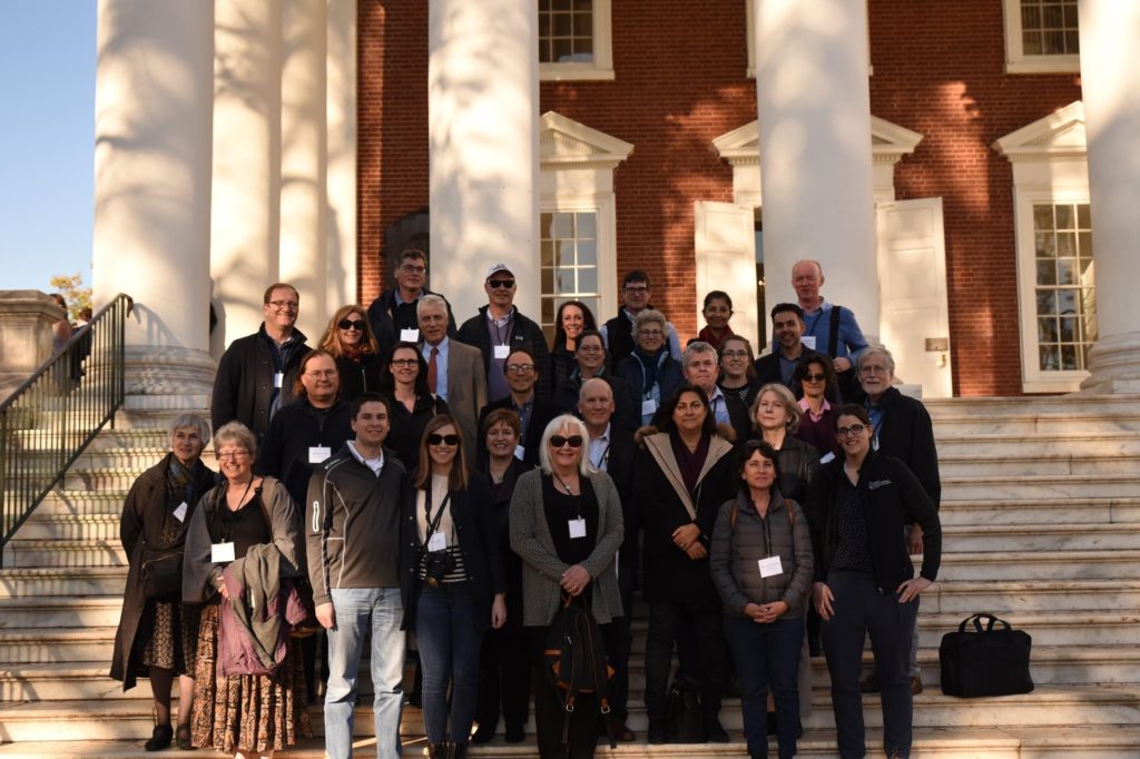
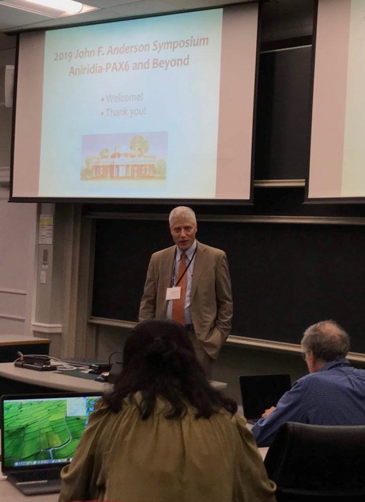
On a crisp Autumn weekend in early November 2019, more than three dozen scientists, clinicians, and patient representatives met on the campus of the University of Virginia to share their work on aniridia-related research and initiatives.
The meeting was unusual in several ways. Instead of a large conference, the small number of participants allowed for in-depth group discussion. Instead of focusing on a single area of research, the agenda spanned a variety of disciplines, which gave participants fresh context and new ideas for their own work. And the meeting also included a number of patients and patient advocacy group representatives, who not only learned more about the research process but also contributed their unique perspectives to the discussion.
Introduction: “Looking back and peering forward”
Veronica van Heyningen
University College, London
Dr van Heyningen spoke about how the work of many researchers is broadening our understanding of the many different types of aniridia-related gene mutations. This work has led to current advances, such as the use of iPSC (induced pluripotent stem cells) to study the cornea problems associated with aniridia. It has also led to important new questions about aniridia-related problems with sleep, obesity, and neurological (brain) issues. In the future, this work could lead to gene therapies, improvements in surgical techniques, and perhaps even to technology such as an “artificial retina.”
In 1992, Veronica van Heyningen was instrumental in the discovery that PAX6 is the primary gene involved with aniridia. Although semi-retired now, Veronica van Heyningen continues to work on several aniridia-related projects, and serves as Patron of Aniridia Network UK

“Phenotype/Genotype correlation in aniridia”
Dominique Bremond-Gignac
Universite Paris V Descartes, Hopital Universitaire Necker, Paris
Visual impairment in aniridia can occur from various mechanisms, such as:
- Glaucoma
- Aniridic keratopathy and limbal cell insufficiency (too few of the cells near the cornea that help it to heal)
- Optic nerve hypoplasia (underdevelopment of the optic nerve)
- Foveal hypoplasia (underdevelopment of the fovea, an area on the retina where vision is sharpest)
Dr. Bremond-Gignac noted that limbal cell insufficiency as the sole cause of aniridic keratopathy has become controversial. Many researchers are now considering that keratopathy in aniridia is likely the result of multiple problems, including abnormalities in the tear film of patients with aniridia.
Dr. Bremond-Gignac is the Chairperson of the Scientific Committee of Aniridia Europe. Aniridia Europe is a member of ERN-EYE, a European Reference Network dedicated to Rare Eye Diseases.
“Using genomics to elucidate mechanisms of acquired ocular disease in aniridia”
Melinda Duncan
University of Delaware
Dr. Duncan discussed cataract and cornea problems in patients with aniridia. Her work investigates why these problems occur. One clue is that the Pax6 gene is expressed (makes a protein) throughout life. In people with aniridia, only 50% of this protein is made. In the lens, Pax6 helps lens cells “remember” to be lens cells. Without it, lens cells appear to make molecules that cause them to act more like scar cells. These molecules may also sensitize the rest of the eye to form scar tissue.
- The epithelium (the outermost layer) of the cornea in the aniridic eye is very fragile, and prone to damage. Limbal cell insufficiency is part of the problem but seems to be just one part.
- Physicians should treat aniridic eyes as more likely to form scar tissue than typical eyes. Surgery should be avoided whenever possible.
“Genotype-phenotype studies of aniridia-associated keratopathy in European cohorts”
Neil Lagali
Linköping University, Sweden
More than 400 unique PAX6 mutations have been reported. There are also additional genes involved in some cases of aniridia. This work involved assessment of 92 eyes for aniridia-associated keratopathy. The researchers graded keratopathy on a scale of 0-4, and compared clinical observations with the patient’s specific mutation.
In this study:
- 60 eyes had confirmed Pax6 mutation/deletion. These patients had a lower age, worse acuity, corneal sensitivity, and epithelium changes than the patients who had aniridia without Pax6 mutation (10 eyes).
- Patients with WAGR syndrome (deletion of Pax6) had the most aggressive keratopathy
The researchers concluded that aniridia-associated keratopathy is likely a condition with multiple subtypes. Some mutation types were clearly associated with more severe presentation and progression. Additional research is needed to help classify where each mutation type falls on the keratopathy spectrum. This research could help doctors diagnose and treat different subtypes of keratopathy and help with future clinical trials.
The results of this study were published by the American Academy of Ophthalmology in the article "PAX6 Mutational Status Determines
Aniridia-Associated Keratopathy Phenotype."
“Anterior Chamber Angle in Aniridia”
Peter Netland
University of Virginia
In the past, it was thought that progressive closure of the anterior chamber angle was responsible for the development of glaucoma in patients with aniridia. This study examined 43 patients (86 eyes) using high-resolution anterior segment optical coherence tomography (OCT) or clinical gonioscopy, or both.
- In this study, most eyes with aniridia and glaucoma had open anterior chamber angles
- The small number of eyes with aniridia and glaucoma that had closed angles all had a prior history of some type of eye surgery
- 50% of aniridia patients have some evidence of glaucoma by 8 years of age
- Prophylactic goniotomy has been proposed as a possible treatment for aniridic eyes that are developing progressive closure of the angle
- “goniotomy is usually not helpful for aniridia patients, although the procedure may be considered in the uncommon situation of an infant with aniridia and glaucoma”
- Surgical approaches that remove tissue from the angle, such as Trabectome, are under evaluation. Often clinicians will treat with glaucoma drainage implant in aniridia patients who develop intractable elevation of intraocular pressure despite medical and surgical therapy
- Aniridia is a “profibrotic” disorder (aniridic eyes are more likely to form excess fibrous connective tissue)
- Surgical treatment should be conservative (surgery only when absolutely necessary)
The results of this study were published under the name "Anterior chamber angle in aniridia with and without glaucoma".
“Clinical conundrums in interpreting results of molecular genetic testing”
Kevin Gregory-Evans
University of British Columbia, Eye Care Centre
Dr Gregory-Evans discussed the difficulties involved in providing a diagnosis for patients whose genetic testing results are highly complex. A lively discussion between clinicians and patients/patient advocacy group representatives followed.
Main clinical takeaways for patients:
- What causes aniridic keratopathy? We used to think the cause was Limbal Cell Deficiency (not enough of the cells near the cornea that help it to heal properly). Now it’s thought that in addition to LCD, there are multiple causes, including abnormalities in the chemical makeup of tears in the aniridic eye as well as other abnormalities that are just being discovered.
- More “basic science” research is needed. If more is known about what actually causes keratopathy, then treatments could be designed that target those abnormalities
- Aniridic corneas are fragile
- Eye drops should be preservative-free whenever possible
- Autologous serum drops may be helpful for keratopathy
- Amniotic membrane does not appear to be as helpful as hoped
- Contact lenses should be avoided
- Aniridic eyes are prone to excess scar formationSurgery should be avoided unless absolutely necessary
- When surgery is required, “less is best” Surgeons should strive for least invasive techniques
- Clinicians at this meeting urged caution with artificial iris implants, citing concerns about complications such as fibrosis, glaucoma